BREAKING NEWS: New drug approved by the FDA for Duchenne muscular dystrophy. Learn more about this new treatment option.

An icon that marks all of our informational disease pages

Congenital Myopathies

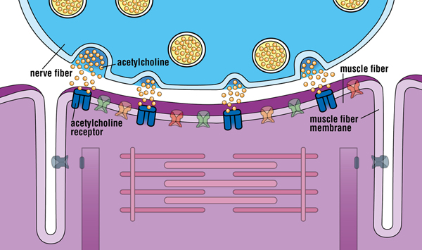
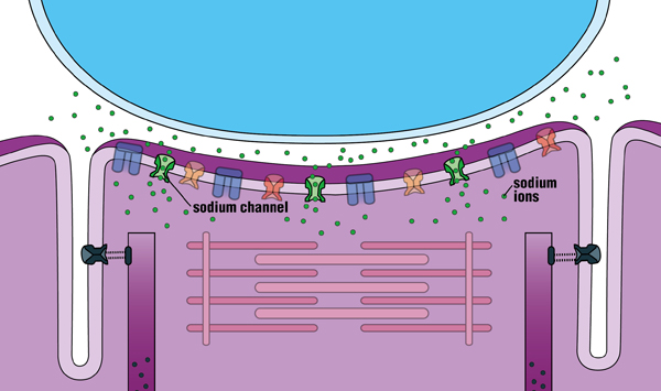
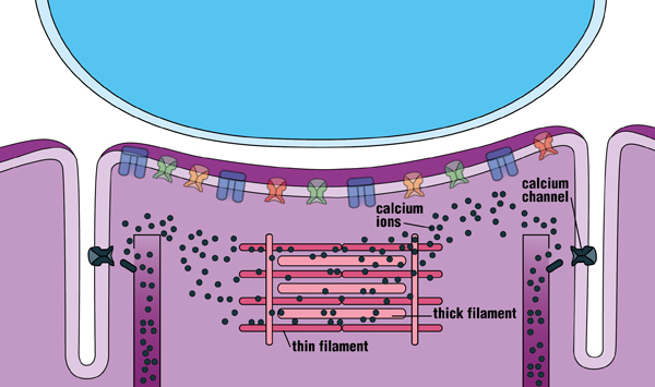
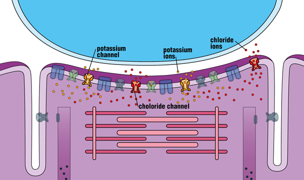
How Ion Channels Regulate Muscle Contraction

Acetylcholine leaves the nerve fiber and docks on receptors in the muscle membrane, causing that area of the muscle fiber to become slightly more positive (“depolarized”).


Sodium channels open in response to this small depolarization, permitting a huge flow of positively charged sodium ions to enter the muscle fiber. The depolarization is greatly amplified, and a brief electrical impulse (“action potential”) spreads throughout the fiber.


Depolarization of the muscle fiber is sensed by calcium channels and triggers the release of calcium ions from internal storage areas. This flood of released internal calcium is the chemical signal that causes the thick and thin filaments of the muscle fiber to slide past each another (contract).


The sodium channels spontaneously close, potassium channels open, and positively charged potassium ions exit the fiber. Chloride channels also stay open, and negatively charged chloride ions enter the fiber. All these actions cause the inside of the fiber to become more negative (“repolarized”). The muscle fiber returns to its resting state, calcium is pumped back into the internal storage vesicles, and the fiber is now ready to accept another surge of positively charged ions in response to stimulation from a nerve fiber. (When several muscle fibers are at rest, a muscle can relax.)


Looking for more information, support or ways to get involved?