Inclusion-Body Myositis (IBM)
Diagnosis
Diagnosing inclusion body myositis (IBM) may require a combination of testing modalities. People with the disease may exhibit:
| Test | Characteristic findings |
|---|---|
| Clinical symptoms |
|
| Clinical exam |
|
| Blood tests |
|
| Myositis autoantibody testing |
|
| Assessment of electrical activity in muscles/nerves |
|
| Magnetic resonance imaging (MRI) |
|
| Muscle biopsy |
|
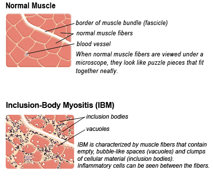
Muscle biopsy is still the gold standard in diagnostic testing for IBM. A biopsy sample from a person with IBM is unique because of its inclusion bodies, for which the disease is named. These “bodies” (which do not appear in normal cells) contain clumps of discarded cellular material. Inflammatory cells can be seen invading muscle tissue, although some researchers believe this invasion is secondary to the primary events in the muscle tissue, presumably those that cause the inclusion bodies to appear.
References
- Inflammatory Muscle Diseases. N. Engl. J. Med. (2015). doi:10.1056/nejmc1506827
- Lotz, B. P., Engel, A. G., Nishino, H., Stevens, J. C. & Litchy, W. J. Inclusion body myositis: Observations IN 40 patients. Brain (1989). doi:10.1093/brain/112.3.727
- Greenberg SA. Inclusion body myositis: clinical features and pathogenesis. Nat Rev Rheumatol. 2019;15(5):257-272. doi:10.1038/s41584-019-0186-x
- Naddaf E. Inclusion body myositis: Update on the diagnostic and therapeutic landscape. Front Neurol. 2022;13:2236. doi:10.3389/FNEUR.2022.1020113/BIBTEX
Last update: Feb 2023
Reviewed by Julie Paik, MD, MHS; Johns Hopkins University
Looking for more information, support or ways to get involved?
Find MDA
in your Community
-

Grants at a Glance
Read More -

Search for Clinical Trials
Learn More

